Biomimetic Therapeutics — Investor Day (March 2008)
37 slides from Biomimetic Therapeutics's investor day presentation, Mar 10, 2008
Slide 1: Welcome to the Analyst and Investor Meeting March 6, 2008
Welcome to the Analyst and Investor Meeting March 6, 2008
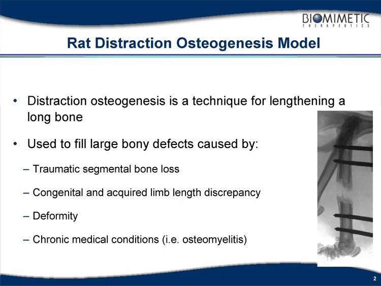
Slide 2: Distraction osteogenesis technique for lengthening a long bone
Distraction osteogenesis is a technique for lengthening a long bone Used to fill large bony defects caused by: Traumatic segmental bone loss Congenital and acquired limb length discrepancy Deformity Chronic medical conditions (i.e. osteomyelitis) Rat Distraction Osteogenesis Model
Slide 3: Study Hypothesis
Study Hypothesis Exogenous rhPDGF-BB will increase new bone formation Rate of healing (bone union) will increase with rhPDGF- BB administration The quantity of new bone formation will increase in a dose dependent manner with rhPDGF-BB
Slide 4: Distraction Osteogenesis Rat Model (RIH)
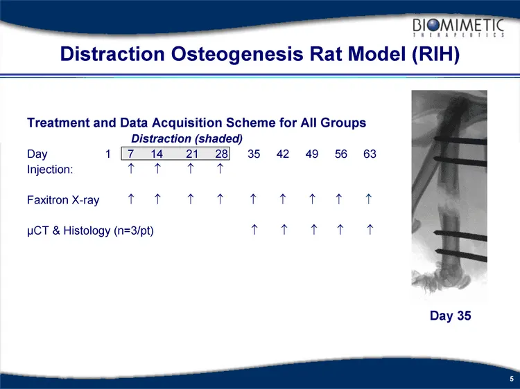
Model Mid-diaphyseal femoral lengthening Seven day latency period 7 mm lengthening over 21 days 2 x 0.17 mm per day Treatments Buffer Injectable Collagen + Buffer Injectable Collagen + rhPDGF-BB (100 ug/ml) Injectable Collagen + rhPDGF-BB (300 ug/ml) Injectable Collagen + rhPDGF-BB (1000 ug/ml) Outc
Slide 5: Treatment and Data Acquisition Scheme for All Groups
Treatment and Data Acquisition Scheme for All Groups Distraction (shaded) Day 1 7 14 21 28 35 42 49 56 63 Injection: ? ? ? ? Faxitron X-ray ? ? ? ? ? ? ? ? ? µCT & Histology (n=3/pt) ? ? ? ? ? Distraction Osteogenesis Rat Model (RIH) Day 35
Slide 6: Progressive Bone Formation and Consolidation
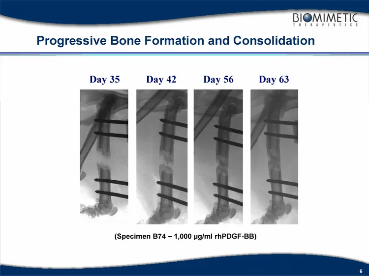
Progressive Bone Formation and Consolidation Day 35 Day 42 Day 56 Day 63 (Specimen B74 - 1,000 µg/ml rhPDGF-BB)
Slide 7: Comparative Evaluation of Treatment Groups
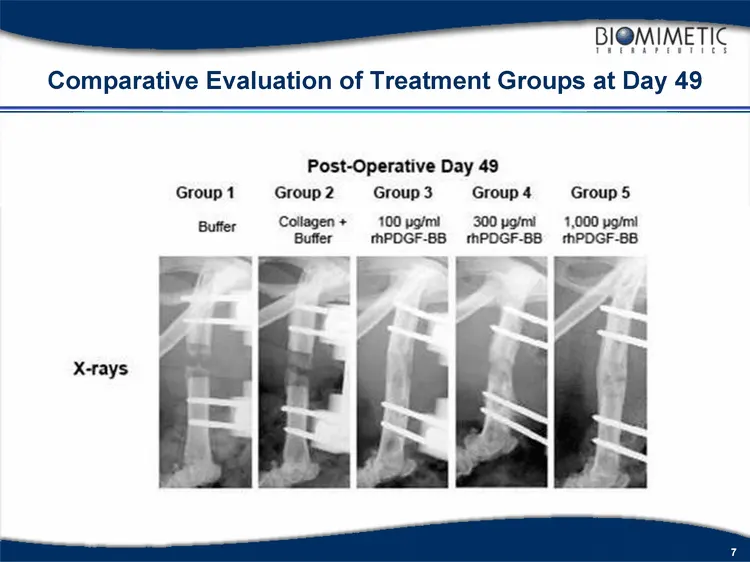
Comparative Evaluation of Treatment Groups at Day 49
Slide 8: Radiographs and µCT images from an animal treated with rhPDGF-BB
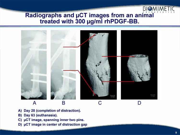
Radiographs and µCT images from an animal treated with 300 µg/ml rhPDGF-BB. A) Day 28 (completion of distraction). B) Day 63 (euthanasia). C) µCT image, spanning inner two pins. D) µCT image in center of distraction gap A B C D
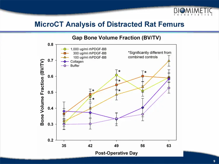
Slide 9: MicroCT Analysis of Distracted Rat Femurs
*Significantly different from combined controls MicroCT Analysis of Distracted Rat Femurs
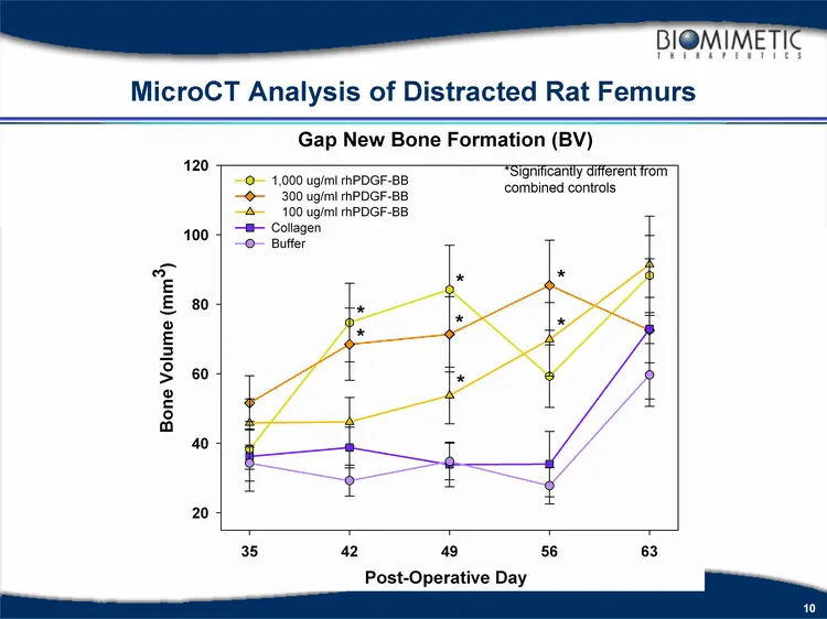
Slide 10: MicroCT Analysis of Distracted Rat Femurs
*Significantly different from combined controls MicroCT Analysis of Distracted Rat Femurs
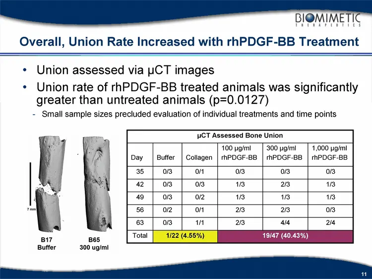
Slide 11: Overall, Union Rate Increased with rhPDGF-BB Treatment
Overall, Union Rate Increased with rhPDGF-BB Treatment µCT Assessed Bone Union µCT Assessed Bone Union µCT Assessed Bone Union µCT Assessed Bone Union µCT Assessed Bone Union µCT Assessed Bone Union Day Buffer Collagen 100 µg/ml rhPDGF-BB 300 µg/ml rhPDGF-BB 1,000 µg/ml rhPDGF-BB 35 0/3 0/1 0/3 0/3
Slide 12: Conclusions
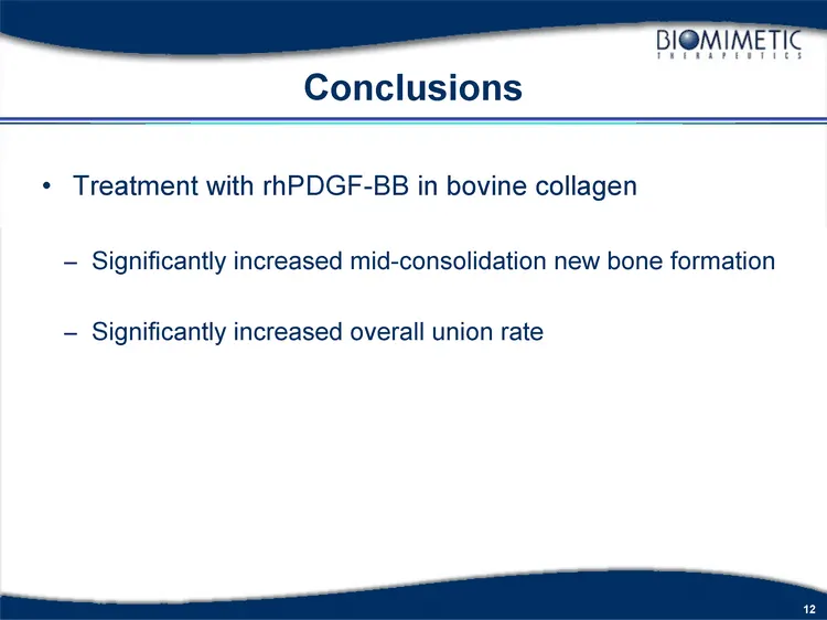
Conclusions Treatment with rhPDGF-BB in bovine collagen Significantly increased mid-consolidation new bone formation Significantly increased overall union rate
Slide 13: Diabetic Rat Model (UMDNJ)
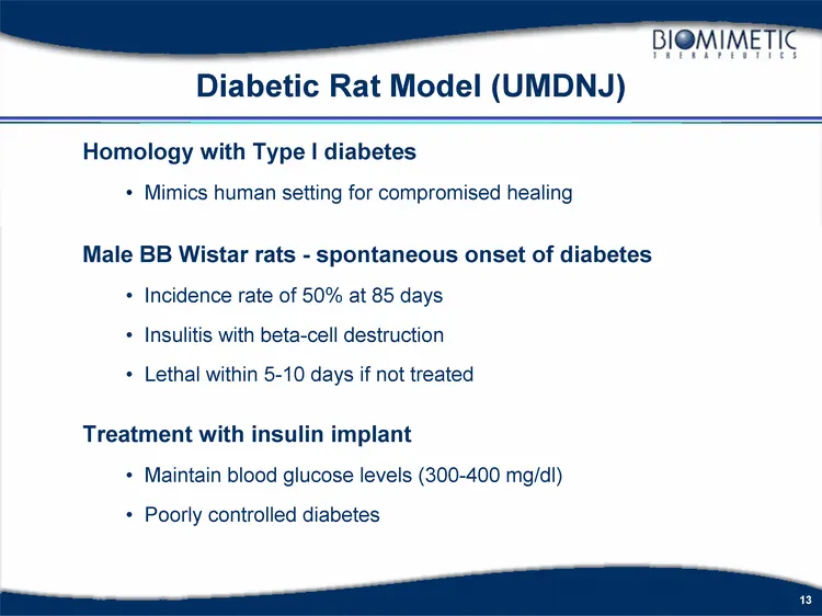
Diabetic Rat Model (UMDNJ) Homology with Type I diabetes Mimics human setting for compromised healing Male BB Wistar rats - spontaneous onset of diabetes Incidence rate of 50% at 85 days Insulitis with beta-cell destruction Lethal within 5-10 days if not treated Treatment with insulin implant Mainta
Slide 14: Diabetic Rat Bone Augmentation Pilot Study
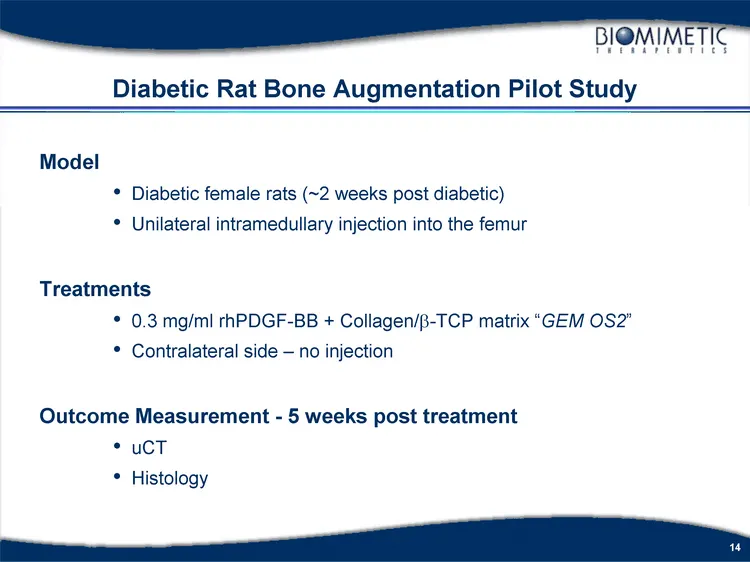
Model Diabetic female rats (~2 weeks post diabetic) Unilateral intramedullary injection into the femur Treatments 0.3 mg/ml rhPDGF-BB + Collagen/b-TCP matrix "GEM OS2" Contralateral side - no injection Outcome Measurement - 5 weeks post treatment uCT Histology Diabetic Rat Bone Augmentation Pilot St
Slide 15: Intramedullary Injection of GEM OS2 Increases Cortical Thickness
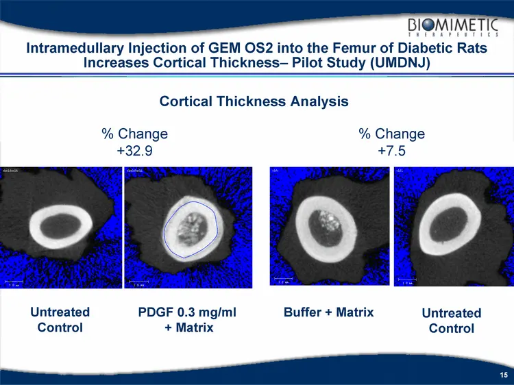
Intramedullary Injection of GEM OS2 into the Femur of Diabetic Rats Increases Cortical Thickness- Pilot Study (UMDNJ) Cortical Thickness Analysis Buffer + Matrix Untreated Control % Change +7.5 Untreated Control % Change +32.9 PDGF 0.3 mg/ml + Matrix
Slide 16: GEMOS2 Injection Causes Formation of Periosteal Woven Bone
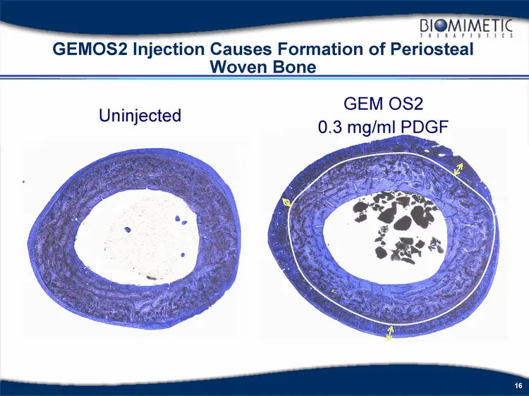
GEMOS2 Injection Causes Formation of Periosteal Woven Bone Uninjected GEM OS2 0.3 mg/ml PDGF
Slide 17: Non-Human Primate Spine Bone Augmentation Study
Non-Human Primate Spine Bone Augmentation Study
Slide 18: Localized Bone Augmentation Concept
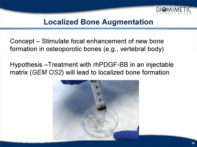
Localized Bone Augmentation Concept - Stimulate focal enhancement of new bone formation in osteoporotic bones (e.g., vertebral body) Hypothesis -Treatment with rhPDGF-BB in an injectable matrix (GEM OS2) will lead to localized bone formation
Slide 19: GEM OS2 Primate Spine Safety Study
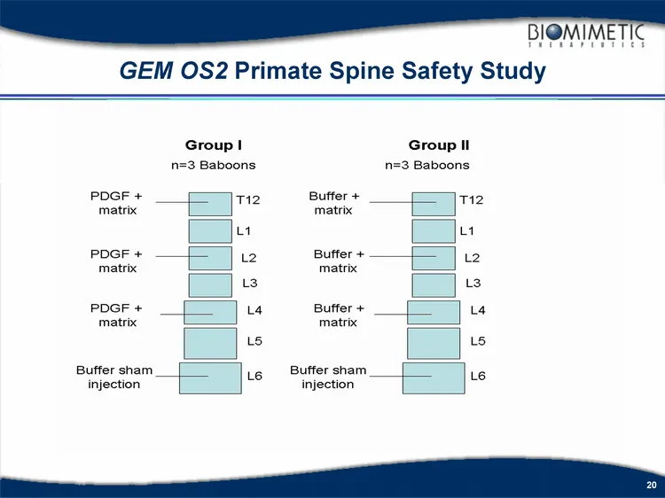
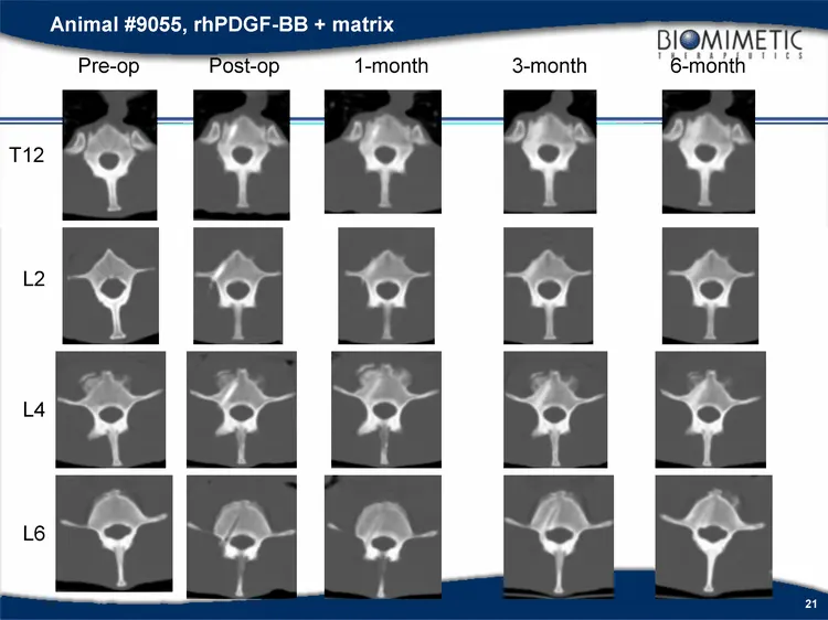
Model Aged female baboons (~17 - 21 years old) Direct injection into the vertebral body Treatments 1.0 mg/ml rhPDGF-BB + Collagen/b-TCP matrix "GEM OS2" Buffer + Collagen/b-TCP matrix Buffer alone Outcome Measurements - pre-op, post-op, 1, 3, 6 & 9 months In-Life Outcomes: MRI, QCT, X-rays, Clinical
Slide 20: GEM OS2 Primate Spine Safety Study
GEM OS2 Primate Spine Safety Study
Slide 21: Animal #9055, rhPDGF-BB + matrix
Animal #9055, rhPDGF-BB + matrix Pre-op Post-op 1-month 3-month 6-month T12 L2 L4 L6
Slide 22: GEM OS2 Injection Increases vBMD Post-surgery
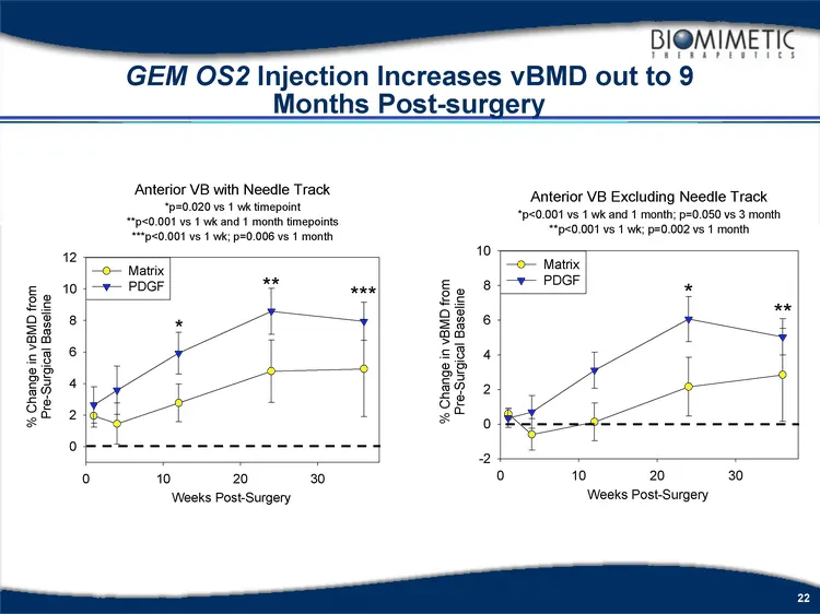
GEM OS2 Injection Increases vBMD out to 9 Months Post-surgery
Slide 23: Summary - Bone Augmentation with rhPDGF-BB
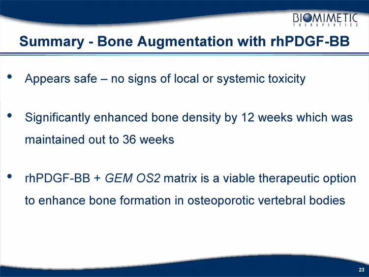
Appears safe - no signs of local or systemic toxicity Significantly enhanced bone density by 12 weeks which was maintained out to 36 weeks rhPDGF-BB + GEM OS2 matrix is a viable therapeutic option to enhance bone formation in osteoporotic vertebral bodies Summary - Bone Augmentation with rhPDGF-BB
Slide 24: Seven Ongoing Trials in Orthopedics
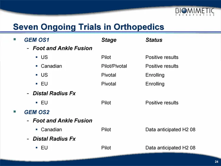
Seven Ongoing Trials in Orthopedics GEM OS1 Stage Status Foot and Ankle Fusion US Pilot Positive results Canadian Pilot/Pivotal Positive results US Pivotal Enrolling EU Pivotal Enrolling Distal Radius Fx EU Pilot Positive results GEM OS2 Foot and Ankle Fusion Canadian Pilot Data anticipated H2 08 Di
Slide 25: US Pivotal GEM OS1 Clinical Trial
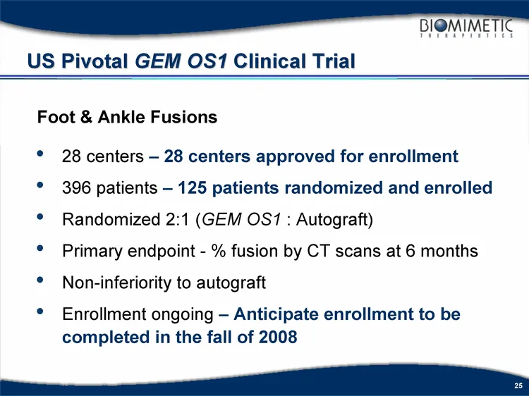
US Pivotal GEM OS1 Clinical Trial 28 centers - 28 centers approved for enrollment 396 patients - 125 patients randomized and enrolled Randomized 2:1 (GEM OS1 : Autograft) Primary endpoint - % fusion by CT scans at 6 months Non-inferiority to autograft Enrollment ongoing - Anticipate enrollment to be
Slide 26: EU Pivotal GEM OS1 Clinical Trial
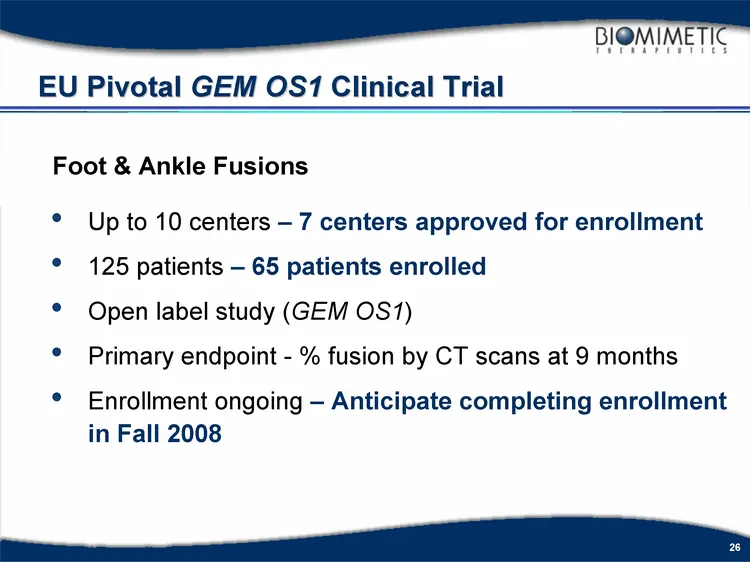
EU Pivotal GEM OS1 Clinical Trial Up to 10 centers - 7 centers approved for enrollment 125 patients - 65 patients enrolled Open label study (GEM OS1) Primary endpoint - % fusion by CT scans at 9 months Enrollment ongoing - Anticipate completing enrollment in Fall 2008 Foot & Ankle Fusions
Slide 27: Canadian GEM OS1 Registration Clinical Trial
Canadian GEM OS1 Registration Clinical Trial 3 centers 60 patients Open label study (GEM OS1) Primary endpoint - % fusion at 9 months Enrollment completed Foot & Ankle Fusions
Slide 28: Canadian GEM OS1 Registration Clinical Trial Filing Status
Canadian GEM OS1 Registration Clinical Trial Filing Status - Based upon ongoing conversations with Health Canada related to the filing of the DLA, the need to hold a face-to-face meeting with BioMimetic has been eliminated. The Company is addressing comments provided by Health Canada and intends to
Slide 29: Summary of N. American Foot and Ankle rhPDGF Clinical Trials
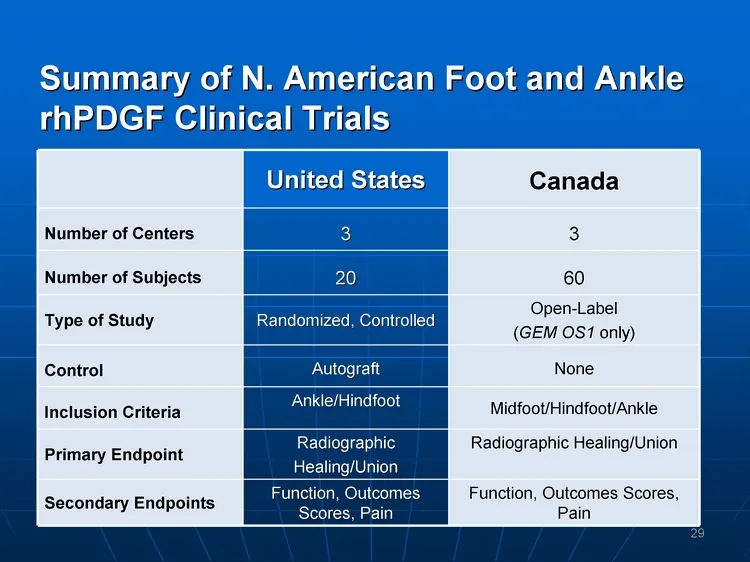
Summary of N. American Foot and Ankle rhPDGF Clinical Trials United States Canada Number of Centers 3 3 Number of Subjects 20 60 Type of Study Randomized, Controlled Open-Label (GEM OS1 only) Control Autograft None Inclusion Criteria Ankle/Hindfoot Midfoot/Hindfoot/Ankle Primary Endpoint Radiographi
Slide 30: Patient Demographics
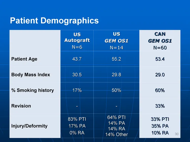
Patient Demographics US Autograft N=6 US GEM OS1 N=14 CAN GEM OS1 N=60 Patient Age 43.7 55.2 53.4 Body Mass Index 30.5 29.8 29.0 % Smoking history 17% 50% 60% Revision - - 33% Injury/Deformity 83% PTI 17% PA 0% RA 64% PTI 14% PA 14% RA 14% Other 33% PTI 35% PA 10% RA 30
Slide 31: Types of Procedures Performed
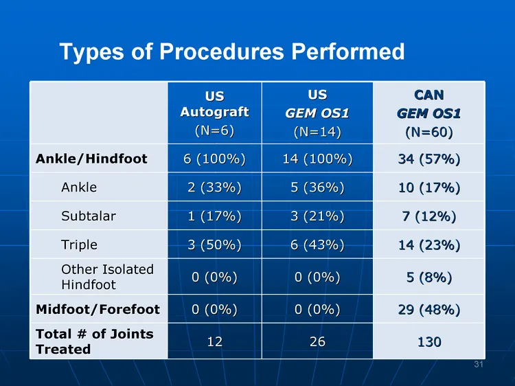
US Autograft (N=6) US GEM OS1 (N=14) CAN GEM OS1 (N=60) Ankle/Hindfoot 6 (100%) 14 (100%) 34 (57%) Ankle 2 (33%) 5 (36%) 10 (17%) Subtalar 1 (17%) 3 (21%) 7 (12%) Triple 3 (50%) 6 (43%) 14 (23%) Other Isolated Hindfoot 0 (0%) 0 (0%) 5 (8%) Midfoot/Forefoot 0 (0%) 0 (0%) 29 (48%) Total # of Joints Tr
Slide 32: Results: Radiographic and Clinical Union
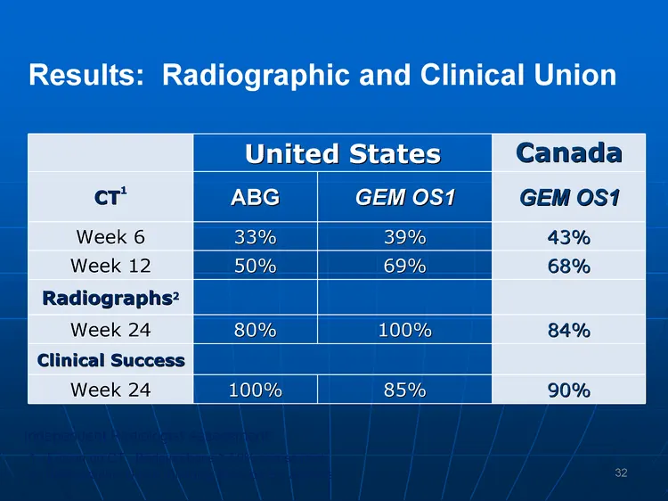
United States United States Canada CT1 ABG GEM OS1 GEM OS1 Week 6 33% 39% 43% Week 12 50% 69% 68% Radiographs2 Week 24 80% 100% 84% Clinical Success Week 24 100% 85% 90% Results: Radiographic and Clinical Union Independent Radiologist Assessment: 1. Fusion on CT: Bridging bone ^ 50% across joint 2.
Slide 33: Results: Safety and AOFAS Scores
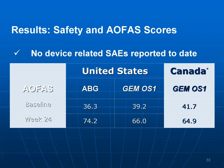
United States United States Canada* AOFAS ABG GEM OS1 GEM OS1 Baseline 36.3 39.2 41.7 Week 24 74.2 66.0 64.9 Results: Safety and AOFAS Scores No device related SAEs reported to date *Hindfoot / ankle only 33
Slide 34: Results: Donor Site Pain and Procedure Time
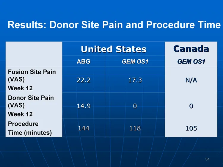
United States United States Canada ABG GEM OS1 GEM OS1 Fusion Site Pain (VAS) Week 12 22.2 17.3 N/A Donor Site Pain (VAS) Week 12 14.9 0 0 Procedure Time (minutes) 144 118 105 Results: Donor Site Pain and Procedure Time 34
Slide 35: Discussion
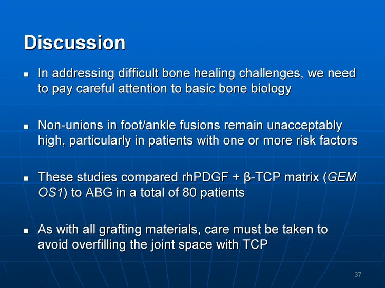
Discussion In addressing difficult bone healing challenges, we need to pay careful attention to basic bone biology Non-unions in foot/ankle fusions remain unacceptably high, particularly in patients with one or more risk factors These studies compared rhPDGF + ^-TCP matrix (GEM OS1) to ABG in a tota
Slide 36: Discussion
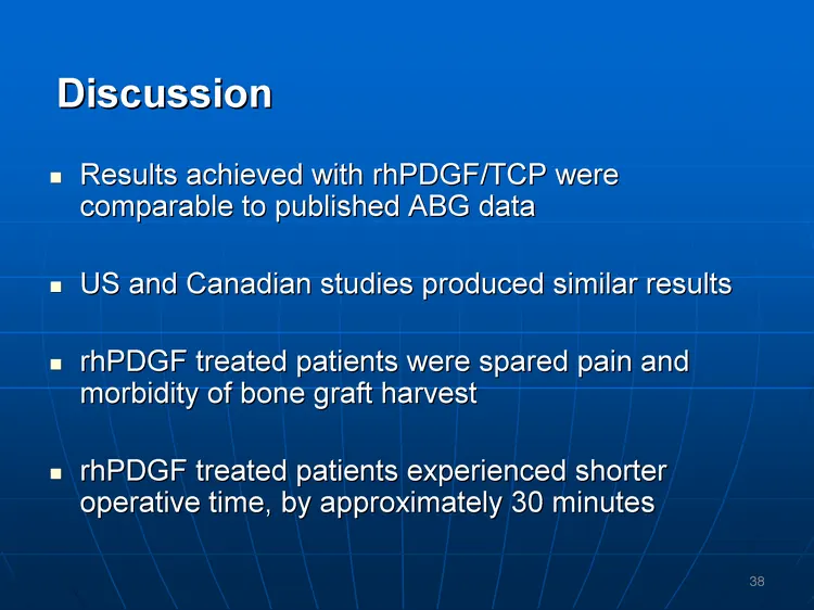
Discussion Results achieved with rhPDGF/TCP were comparable to published ABG data US and Canadian studies produced similar results rhPDGF treated patients were spared pain and morbidity of bone graft harvest rhPDGF treated patients experienced shorter operative time, by approximately 30 minutes 38
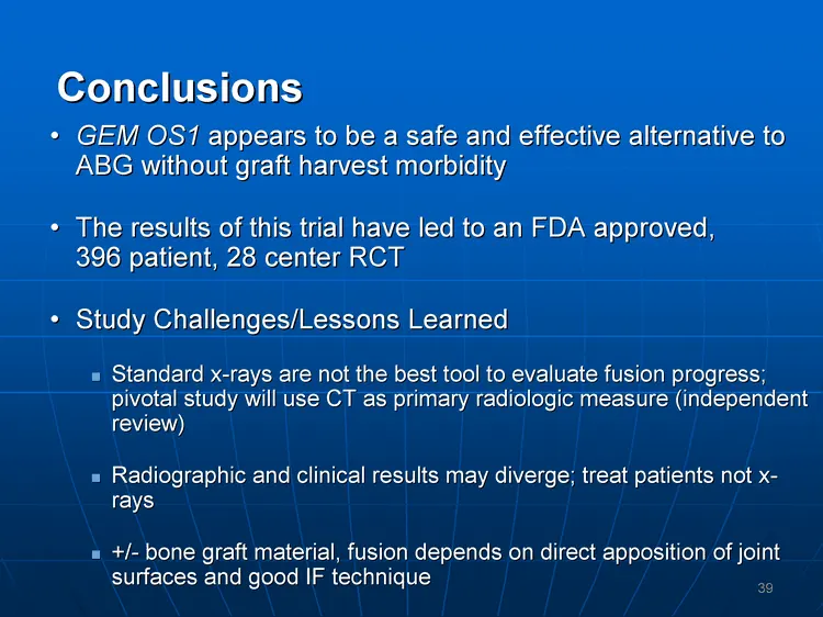
Slide 37: Conclusions
Conclusions GEM OS1 appears to be a safe and effective alternative to ABG without graft harvest morbidity The results of this trial have led to an FDA approved, 396 patient, 28 center RCT Study Challenges/Lessons Learned Standard x-rays are not the best tool to evaluate fusion progress; pivotal stud
Source: SEC EDGAR · 8-K filed Mar 10, 2008
More decks from Biomimetic Therapeutics Inc
 31st Annual J.P. Morgan Healthcare Conference 201341 slides · Jan 8, 2013
 Technical Presentation (June 2012)26 slides · Jun 21, 2012
 Jefferies Global Healthcare Conference 201136 slides · Jun 10, 2011
 Investor Day (February 2011)113 slides · Feb 23, 2011
 2010 AOFAS Annual Summer Meeting19 slides · Jul 7, 2010
 Technical Presentation (July 2010)19 slides · Jul 7, 2010